

A developmental atlas of the Arabidopsis ovule from Vijayan et al. 2021, eLife.

Cells from the surface of an imaginal wing disc of Drosophila, colored by area. The peripodial membrane (green) was digitally removed from the confocal stack in MorphoGraphX prior to surface extraction. Data from Aegerter-Wilmsen et al. 2012, Development.
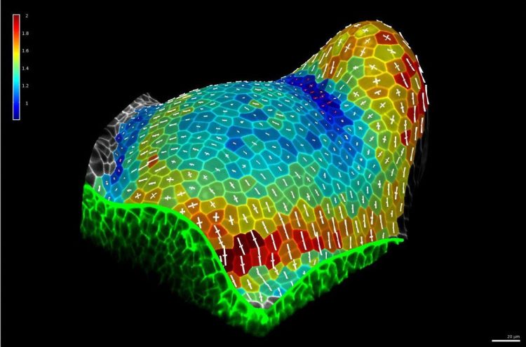
Principal directions of growth and areal expansion of cells in a tomato shoot apex, extracted from time-lapse confocal microscopy data from Kierzkowski et al. 2012, Science.
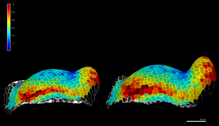
Side views of a growing tomato shoot apex, over 11h time interval. Cells colored by areal expansion. Data from Kierzkowski et al. 2012, Science.

Full 3D segmentation of an Arabidopsis thaliana mature embryo. Cells colored by ID. Data from Bassel et al. 2014, PNAS.
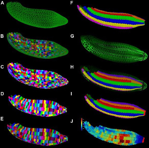
Workflow of 3D segmentation and cell classification in an Arabidopsis thaliana mature embryo. Published in Bassel et al. 2014, PNAS.

Mechanical simulation on a 3D segmentation of an Arabidopsis thaliana mature embryo. Published in Bassel et al. 2014, PNAS.
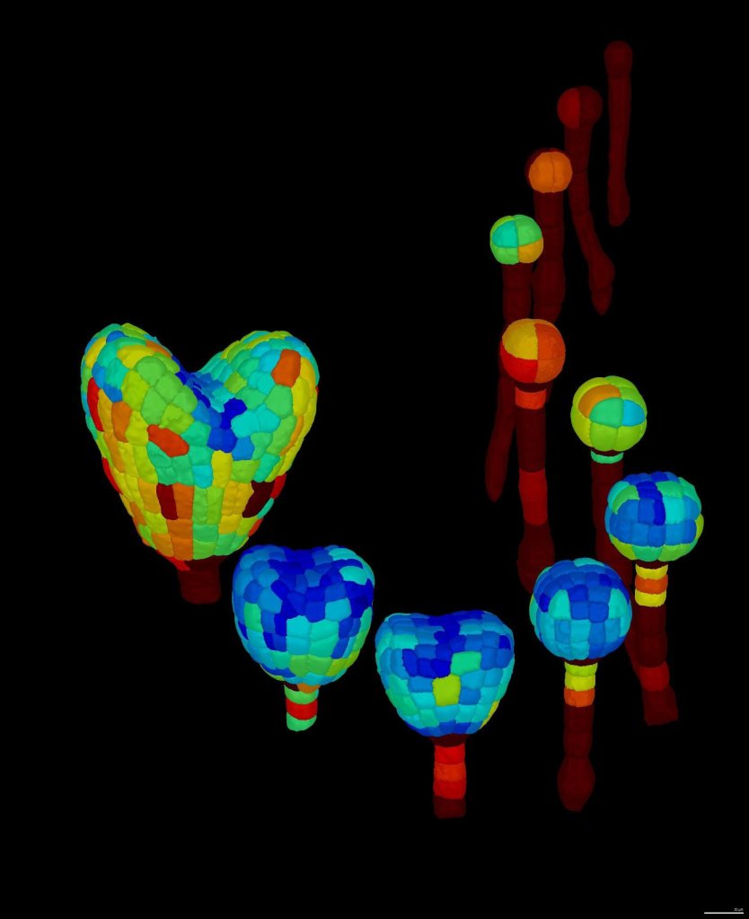
Full 3D segmentation of Arabidopsis thaliana embryos at different developmental stages. Cells colored by volume. Published in Yoshida et al. 2014, Developmental cell.
Videos
For more videos, please see our YouTube channel.
